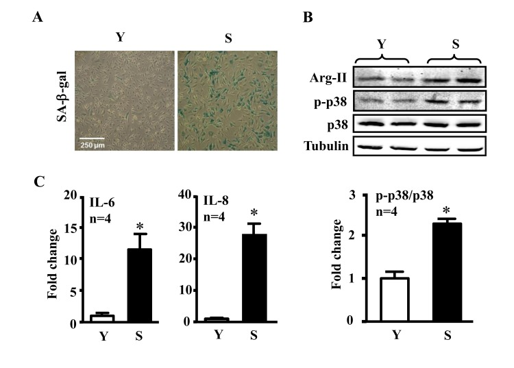
Figure 1.Comparison of inflammatory cytokines between young and senescent endothelial cellsConditioned medium and cell lysate were collected from young (Y) and senescent (S) HUVECs which were serum-starved overnight. (A) SA-β-gal staining. (B) Immunoblotting to detect Arg-II, phsopho-p38-Thr180/Tyr182 (p-p38) and total-p38 (p38) in young and senescent cells. Tubulin served as loading control. (C) The secretion of IL-6 and IL-8 was evaluated by ELISA with collected conditioned medium described above. n=4, *p<0.05 vs young cells (Y).