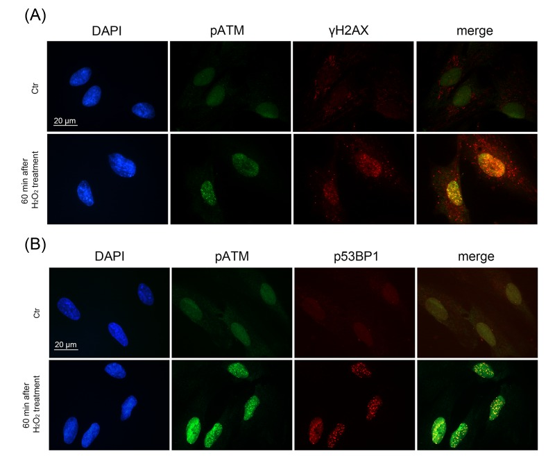
Figure 3.Formation of DNA damage foci containing pATM, γH2AX, p53BP1 in H2O2-treated hMESCsImmunofluorescent analysis with the use of specific antibodies against pATM, γH2AX, p53BP1 revealed co-localization of pATM with either γH2AX (A) or p53BP1 (B) in 60 min after beginning of H2O2 treatment. DAPI was used as nuclear stain (blue). Images are taken at magnification X100. Scale bar is 20 µm and valid for all images.