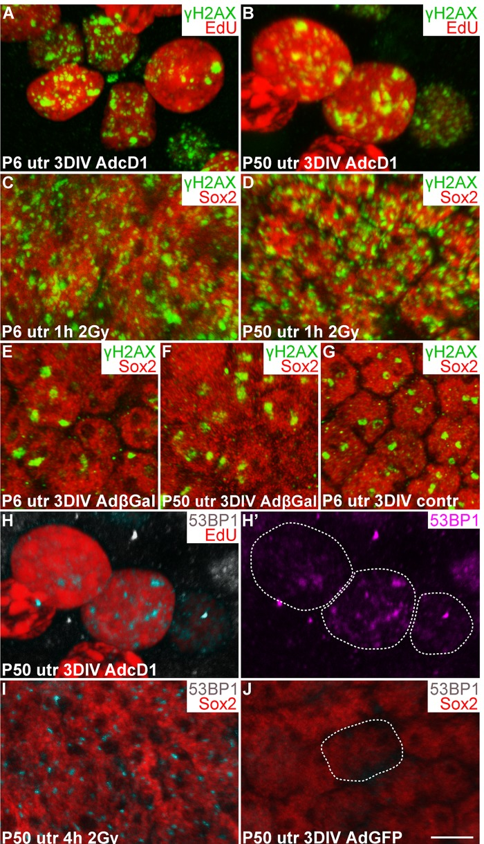
Figure 3.Utricular supporting cells show γH2AX and 53BP1 foci upon forced cell cycle re-entry, as revealed by confocal imaging. (A,B) At 3 DIV, AdcD1-infected P6 and P50 utricles display EdU+ SCs with numerous, small γH2AX foci. (C,D) Similar γH2AX profiles are seen 1 h post-irradiation in Sox2+ utricular SCs of both ages. (E-G) At 3 DIV, Sox2+ SCs in both AdβGal-infected P6 and P50 utricles, and in non-infected P6 utricle show one or two large γH2AX foci per nucleus. (H,H') At 3 DIV, EdU+ SCs in AdcD1-infected P50 utricle display 53BP1 foci. Edu+ SC nuclei containing 53BP1 foci are outlined (H'). (I) Four hours post-irradiation, P50 utricular Sox2+ SCs show 53BP1 foci. (J) At 3 DIV, AdGFP-infected P50 utricle is devoid of 53BP1 foci. A Sox2+/GFP+/53BP1- nucleus is outlined. Abbreviations: AdcD1, adenovirus encoding cyclin D1; AdβGal, adenovirus encoding β-galactosidase; AdGFP, adenovirus encoding green fluorescent protein; Gy, gray; γH2AX, Ser 139 phosphorylated histone H2AX; utr, utricle. Scale bar, shown in J: A-J, 5 µm.