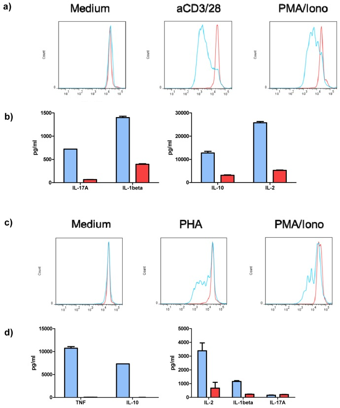
Figure 3.VGF induces proliferation and cytokine production by T cells and PBMCsIsolated T cells were stimulated with aCD3/28 or PMA/Ionomycin (as positive control) or were left untreated (medium control) for 3 days in the presence of anti-VGF (D20; red) or an isotype-matched control Ab (blue). Division of cells is detected by loss of fluorescence intensity of cells labeled with CFSE. Blockade of VGF inhibited the proliferation of T cells stimulated with aCD3/28 or PMA/Ionomycin (a) and reduced the production of cytokines in T cell cultures stimulated with aCD3/28 (b). Blocking of VGF diminished the division of PBMCs stimulated with PHA or PMA/Ionomycin (as positive control; c) and also the secretion of cytokines in PHA-stimulated PBMC cultures.