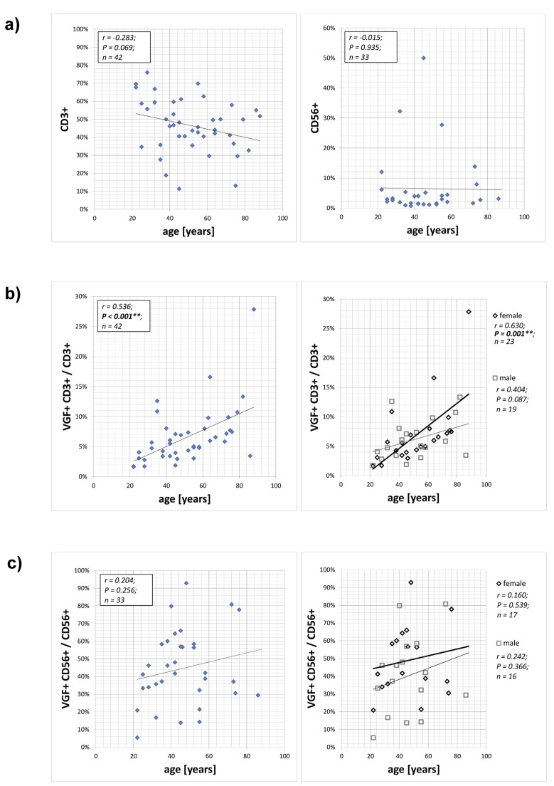
Figure 1.Expression of VGF on CD3+ T cells and CD56+ NK cellsPBMCs isolated from 42 healthy volunteers aged between 22 and 88 years without psychiatric diagnosis were stained with anti-VGF (D20) and anti-CD3 or anti-CD56 and were analyzed using flow cytometry. The numbers of CD3+ T cells and CD56+ NK cells depending on the age of the persons is shown in (a). The frequency of VGF-expressing CD3+ cells within the T lymphocytes population is correlated with the age of the cohort (b; left) and further subdivided according to the gender (b; right). The proportion of VGF-expressing CD56+ cells within the NK cell population is shown in dependence with the age of the cohort (c; left) and further subdivided according to the gender (c; right).