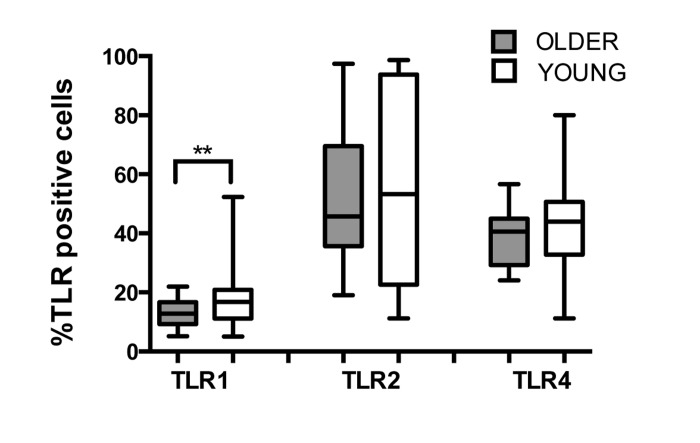
Figure 1.Effect of aging on expression of TLRs in human PMNWhole blood of younger (n=31) and older (n =22) adults was labeled for flow cytometry with lineage markers and TLRs at 4°C for 30 min following RBC lysis. Labeling was detected by LSR II. Data shown are % positive neutrophils for TLR surface expression. Values indicate the means ± SEM in young and older adults. Asterisks indicate statistical significance between younger and older cohort (Unadjusted t-test accounting for unequal variances, p < 0.02).