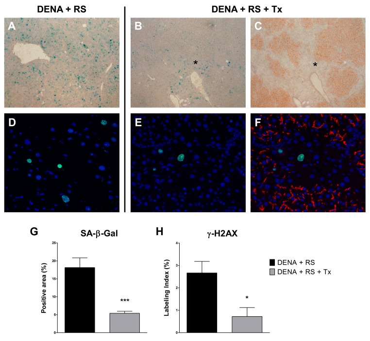
Figure 3.Hepatocyte transplantation reverses the RS-induced senescent phenotypeExpression of SA-β-gal (panels A, B, C and G) and γ-H2AX (panels D, E, F and H), in rat liver exposed to either DENA+RS or DENA+RS followed by normal hepatocyte transplantations. Markers of cell senescence were highly expressed in DENA+RS-treated livers (panels A, D), while their levels were markedly reduced in animals receiving hepatocyte transplantation (panels B, C, E, F). In the latter group, extensive repopulation of the recipient liver was observed (panels C, histochemical staining for DPP-IV, orange-rust; panel F, immunofluorescence staining for CD26, red); note the residual expression of senescence markers in non-repopulated areas (panel C and F). Panels A, B and C: magnification 40x; panels D, E and F: magnification 200x. Panels G and H: ***P<0.001; *P<0.05.