Submit an Article
Navigate
Home
Editorial Board
Editorial Policies
Current Volume
Archive
Scientific Integrity
Publication Ethics Statements
Interviews with Outstanding Authors
Newsroom
Sponsored Conferences
Podcast
Contact
Special Collections
Submit an Article
Online ISSN: 1945-4589
Research Paper
|
Volume 5, Issue 6
|
pp. 474–484
Rapamycin suppresses brain aging in senescence-accelerated OXYS rats
Back to article
Figure 6
(6 of 6)
−
100%
+
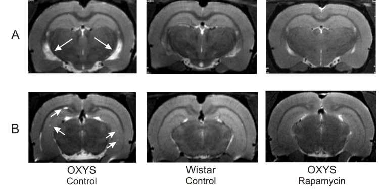
Figure 6.
MRI image of brain 4 month-old OXYS, Wistar rats and OXYS rats after rapamycin supplementation. (
A
) Hydrocephaly of lateral ventricle of OXYS rats (arrow). (
B
) Loci of demyelinization of the brain OXYS rats (arrow).