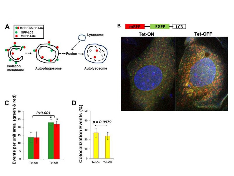
Figure 6.Increased autophagy in Tet-OFF MHEC using adenovirus expressing mRFP-GFP-LC3(A) Schematic presentation shows that GFP-LC3 (green) and mRFP-LC3 (red) signals are present on the autophagosome, whereas autolysosome contains only mRFP-LC3 (red) signals [40]. (B) Confocal microscopy of Adv-mRFP-GFP-LC3-transduced MHECs. (C) Quantification of red and green fluorophores using NIH ImageJ 1.47b demonstrate >1.5-fold increase in autophagy in Tet-OFF MHEC (n=50 cells). (D) Quantification of colocalization events (yellow) using spatial overlap of red and green was done in Fiji program (ImageJ 1.47h). 1.5-fold increase in overall autophagy in Tet-OFF MHEC (C), but no significant changes in the ratio of intracellular red vs. green in Tet-OFF MHEC (C), and no changes in the colocalization signals between Tet-ON vs. Tet-OFF (D) suggest an effective autophagic flux in Tet-OFF MHEC. *p<0.05.
Figure 6 — Oxidative stress improves coronary endothelial function through activation of the pro-survival kinase AMPK | Aging