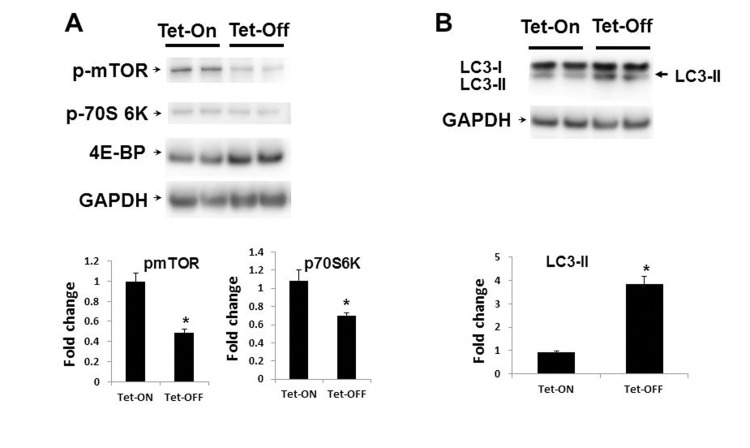
Figure 5.Inhibition of mTOR signaling in Tet-OFF MHEC with high ROS(A) Western blot analyses of MHEC protein lysates from two independent lines of NVF Tet-ON and Tet-OFF mice as indicated. WB was carried out using anti-p-mTOR (p-mTOR), anti-p-70S 6k, and anti-4E-BP antibodies. GAPDH was used for loading control. Lower panels, bar graphs show quantitative densitometric analysis of three independent experiments of the p-mTOR and p-70S 6K bands (-fold change expressed in mean ± S.E.M.). *p<0.05 was considered statistically significant. (B) WB analyses of MHEC from two independent lines of NVF Tet-ON and Tet-OFF as in (A) except anti-LC3A (I and II) and ant-GAPDH antibodies were used. Arrow, induction of the autophagy marker LC3-II in Tet-OFF MHEC is indicated. Lower panel, bar graph showing quantitative analyses of LC3-II as indicated. *p<0.05.