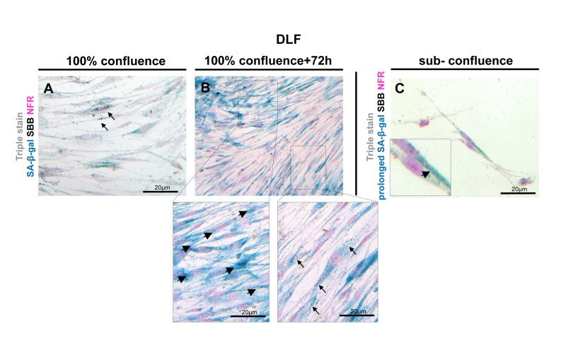
Figure 10.Lipofuscin staining and Senescence-Associated beta-galactosidase (SA-β-gal) activity in primary human diploid lung fibroblasts (DLF)Triple staining of early passage (6th) DLF cells with Sudan Black B (SBB) (dark blue-black granules), SA-β-gal (turquoise color) and Nuclear Fast Red (NFR), as counterstain. (A) Cells that had just reached 100% confluence showed SA-β-gal activity with negligible lipofuscin (arrows). (B) In the same assay 72 hours later, some cells demonstrated SA-β-gal staining (arrowheads) without lipofuscin, and there were also cells with concurrent SA-β-gal and SBB staining (arrows). (C) In sub-confluent DLF cells, prolonged SA-β-gal incubation (72 hours) followed by immediate SBB staining, demonstrated SA-β-gal activity (inset) without lipofuscin appearance.