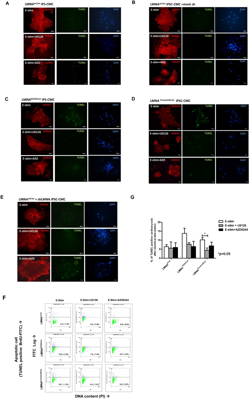
Figure 6.Electrical stimulation inducing apoptosis in cardiomyocytes derived from LMNAR225X/WT &LMNAFrameshift/WT iPSCsRepresentative TUNEL assay and co-immunofluorescence staining of alpha-actinin in cardiomyocytes derived from (A)LMNAWT/WT iPSCs. (B)LMNAR225X/WT iPSCs. (C)LMNAFrameshift/WT iPSCs. (D)LMNAWT/WT iPSCs treated with mock shRNA. (E)LMNAWT/WT iPSCs treated with shLMNA at baseline and after electrical stimulation. (F, G) Quantification of apoptotic cardiac differentiated iPSCs by APO-BrdU TUNEL assay. The percentage of cardiomyocytes with apoptosis was determined by FACS analysis by FL-1 positive gating. Unpaired t-test was performed between treatment and baseline n=3-5, *p-value<0.05.