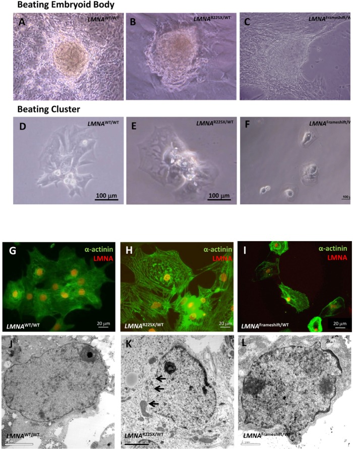
Figure 4.Cardiomyocytes derived from LMNAR225X/WT, LMNAFrameshift/WT and LMNAWT/WT iPSCs(A, B & C) Beating embryoid bodies derived from LMNAR225X/WT, LMNAFrameshift/WT and LMNAWT/WT iPSCs;(D, E & F) After micro-surgical dissection, spontaneously beating cell clusters were plated onto glass coverslips. Videos of these beating embryoid bodies and clusters were available in supplemental materials. (G, H & I) Immunofluorescence co-staining showing the expression of cardiac specific marker, α-actinin and lamin A/C in these cells, (J, K & L) Electronic microscopy of nucleus of cardiomyocytes derived from LMNAR225X/WT, LMNAFrameshift/WT and LMNAWT/WT iPSC line. The black arrows indicate the loss of nuclear envelope in the LMNAR225X/WT iPSC-derived cardiomyocytes.