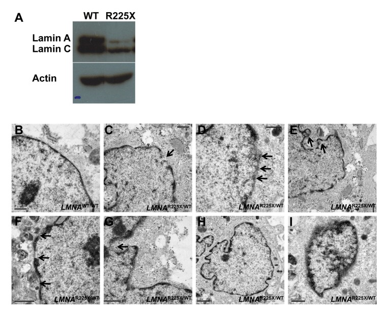
Figure 2.LMNAR225X/WT dermal fibroblasts showing nuclear defects and accelerated apoptosis upon electrical stimulation(A) The expressions of lamin A/C proteins in control and LMNAR225X/WT dermal fibroblasts with western blot analysis using anti-LMNA antibody targeting both N-terminus. (B-I) Electronic micrographs of nuclei of cultured dermal fibroblasts from controls and LMNAR225X/WT: (B) Normal nuclear envelope in control dermal fibroblast;(C) Focal loss of the nuclear membrane in LMNAR225X/WT dermal fibroblasts (arrow);(D) Clustering of nuclear pores;(E, F & G) Bleb and micronucleus formation; (H) Accumulation of mitochondria around the nuclear envelope; (H & I) Irregular shape nucleus, and condensed chromatin.