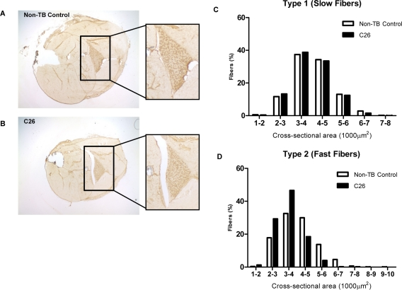
Figure 1.Features of the C26 model of cachexiaFrozen sections of lower hindlimbs of (A) non-tumor-bearing (non-TB) control and (B) C26-bearing mice were immunohistochemically stained for MHC type I protein. Positive myofibers stained a relative dark brown compared to myofibers negative for the protein of interest. (C-D) The whole soleus muscle was subjected to myofiber cross-sectional area analysis to compare the proportion of myofiber type 1 (positively stained) and type 2 (negatively stained) in tumor-bearing mice versus non-TB controls. A more prominent reduction of bigger myofibers and a corresponding increase of smaller myofibers was seen in type 2 myofibers (n = 3).