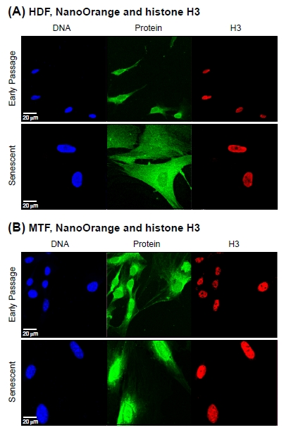
Figure 3.Co-staining of total protein and histone H3Cell we stained with NanoOrange (NO) and subsequently processed for immunofluorescent detection of histone H3 (H3) as indicated in Methods. (A) Representative images acquired with cultures of HDF. The panels are arranged and labeled as indicated in Figure 2A. (B) Representative images acquired with cultures of MTF. The panels are arranged and labeled as indicated in (A) above.