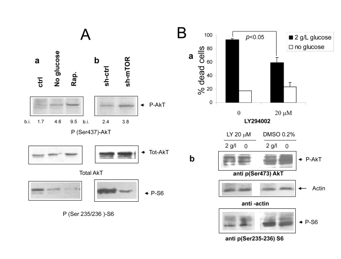
Figure 5. (Aa Immunoblot analysis revealing increased phosphorylation of
AkT/PKB on serine 473 under
nutrient deprivation (upper panel). The relevant band is indicated by the
arrow. Band quantization values (band volume) in band intensity (b.i.)
units are indicated. The same filter was stripped and re-hybridized with an
anti total AkT antiserum to ensure equal protein expression and sample
loading (central panel); a lower strip of the same filter was hybridized
with an antiserum specific for phospho S6 (lower panel, band indicated by
arrow). Picture representative of several independent experiments. b
Protein lysates from mock and mTOR-silenced cells grown under serum free
DMEM with glucose and aminoacids were treated as in A. Relevant bands are
indicated by arrows. Densitometry of p-AkT bands is reported. (B) a
Effect of the PI3 Kinase inhibitor compound LY294402 on Phoenix cell
survival in serum-free medium. Cells were incubated for 72 hours with or without
glucose as indicated. The inhibitor or vehicle alone (DMSO, 1:500 final
dilution) were added at time 0. Values are Mean ±SD of triplicate wells.
Representative of three independent experiments. Note that lower
concentrations of LY294002 had no effect on cell survival in either medium.
b Immunoblot analysis of protein lysates from cells treated
as in a and incubated for 24 hours. Phospho-AkT (serine 473) and phospho-S6
(serine 235-236) were detected by specific antisera. Relevant bands (the
middle one within the triplet for AkT) are indicated by arrows; equal
protein loading was verified by reversible Ponceau S staining.