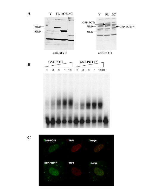
Figure 1.Localization and DNA binding activity of the POT1 ∆C allele.(A) Expression levels of MYC-or GFP-tagged
alleles in HTC75 cells. The full-length (FL, 71kD), POT1∆OB
(∆OB, MW 57kD) and POT1∆C(∆C, MW 43kD) are shown along a
vector-only control. Blots probed with the 9E10 (anti-MYC) (left) or 978 (anti-POT1)
antibodies (right) are shown. (B) Gel shift assay for GST-POT1
and GST-POT1∆C. A 32P-labelled oligonucleo-tide containing
the POT1 minimal binding site was incubated with the amounts of GST fusion
protein shown on top. The free probe is visible at the bottom of the autoradiogram.
(C) Intranuclear localization of GFP-NLS-POT1 and
GFP-NLS-POT1∆C. The GFP-tagged protein is detected in the FITC
channel (left), and telomeres are stained with an anti-TRF1 antibody (371,
middle panels). The overlay is shown in the right panels.