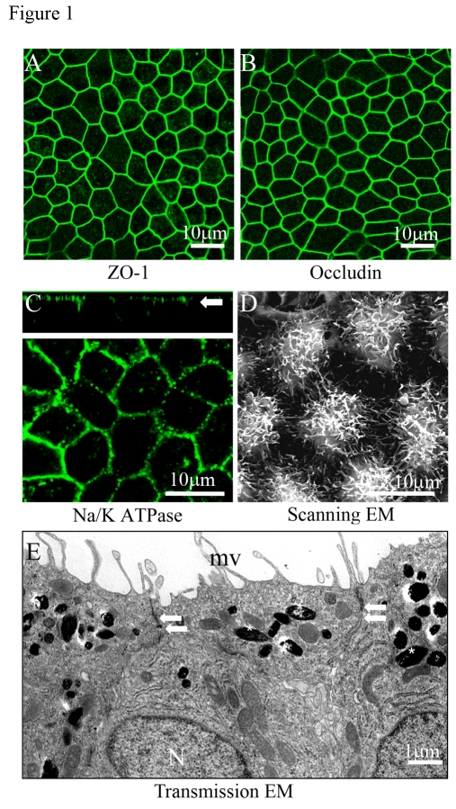
Figure 1.Confocal and electron microscopic characterization of polarized RPE cells.
Evidence for tight junction proteins and polarity in fetal RPE cells
cultured on Transwell filters for 6 weeks. (A, B)
Immunofluorescence staining of tight junction proteins ZO-1 and occludin. (C)
Localization of Na/K- ATPase to the apical plasma membrane as shown in the
confocal vertical (X-Z) section (white arrow). (D) Well
differentiated apical microvilli observed by scanning electron microscopy
(SEM). (E) Well developed microvilli (mv), localization of pigment
on the apical side (asterisks), nuclei on basal side (N), and presence of
tight-junctional complexes (arrows) by transmission electron microscopy
(TEM).