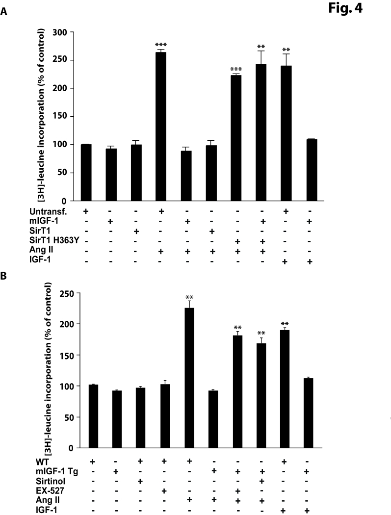
Figure 4.mIGF-1 prevents Ang II- and IGF-1-induced cell hypertrophy ([3H]-leucine incorporation) in HL-1 cardiomyocytes and in mouse neonatal primary cardiomyocytes.(A) HL-1 cardiomyocytes were
transfected with the indicated plasmids, or treated with 20 ng/ml IGF-1 for
24 h, or exposed to Ang II (1 μM
for 24 h). Untransfected cells were used as control (CTL).
Together with Ang II, HL-1 cells were also incubated with 1μCi/ml of [3H]-labeled
leucine (24 h). (B)
Neonatal primary cardiomyocytes from wild type or mIGF-1 Tg mice were
treated with SirT1 inhibitors (sirtinol, 100 μM; EX-527, 1 μM),
or treated with 20 ng/ml IGF-1 for 24 h, or exposed to Ang II (1 μM for 24 h); concomitantly to Ang
II addition, cells were incubated with 1mCi/ml of [3H]-labeled
leucine (24
h).
(A, B) [3H]-leucine incorporation values were normalized to total
protein content and expressed as % of control. Results are means ± SE of 3 independent experiments
(**,***p versus unstimulated
control cells or untreated WT cardiomyocytes).