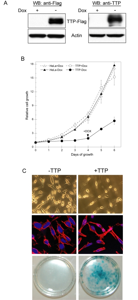
Figure 1.TTP inhibits HeLa cell proliferation through induction of senescence.(A) HeLa
Tet-Off/TTP-Flag cells grown in the presence or absence of 2 μg/ml Dox for
48 hr. The expression of TTP-Flag was detected by western blot (WB) using
antibodies against the Flag epitope (left panel) or TTP (right panel).
Actin was used as a loading control. (B) Growth curves of HeLa
Tet-Off/TTP-Flag (circles) and parental HeLa Tet-Off (triangles) cells in
the presence (open symbols) or absence (filled symbols) of 2 μg/ml Dox. On
day 4 of growth, Dox was added to HeLa Tet-Off/TTP-Flag cells to repress
TTP expression. Each point represents the mean of 4 replicates. (C)
HeLa Tet-Off/TTP-Flag cells were grown in the presence or absence of Dox to
repress (- TTP) or induce (+ TTP) TTP, respectively. Phase contrast (top
panels) and fluorescence (middle panels) microscopy of cells after 48 hr of
TTP expression; original magnification 200X and
400X, respectively. Nuclei (blue) and cytoskeleton (red) are shown in
fluorescent micrographs. HeLa-Tet-Off/TTP-Flag cells were stained for
SA-β-gal activity (bottom panels) after 12 days of TTP expression.