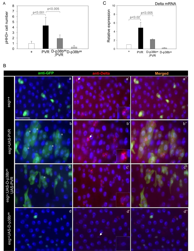
Figure 6.Effects of p38b MAPK knockdown in ISCs and EBs on PVR-induced phenotypes.
(A) Effect of D-p38b knockdown in ISCs/EBs on ectopic PVR-induced
ISC proliferation. The PH3-positive cells in the midgut of the 3-day-old
flies were counted. White bar, esg>+; black bar, esg>UAS-PVR;
dark gray bar, esg>UAS-D-p38bas;UAS-PVR; gray
bar, esg>UAS-D-p38bas. The number of PH3-positive
cells detected per midgut of esg>+ flies was set as 1. P-values
were calculated using Student's t-test. (B) Effect of D-p38b
knockdown in ISCs and EBs on PVR-induced accumulation of large esg- and
Delta-positive cells. The guts of 3-day-old flies were labeled with
anti-Delta and anti-GFP. (a-a''), esg>+; (b-b''), esg>UAS-PVR;
(c-c''), esg>UAS-D-p38bas;UAS-PVR; (d-d''), esg>UAS-D-p38bas.
Overlay (DAPI, blue; anti-Delta, red; anti-GFP, green). Asterisk indicates
EC-like large esg-positive cell. Arrow indicates Delta-positive cells.
Scale bar, 5 μM. C. Effect of
D-p38b knockdown in ISCs and EBs on PVR-induced Delta mRNA expression.
Expression of Delta was measured by quantitative RT-PCR of dissected gut
from 3-day-old flies. White bar, esg>+; black bar, esg>UAS-PVR;
dark gray bar, esg>UAS-D-p38bas;UAS-PVR; gray
bar, esg>UAS-D-p38bas. Expression was normalized to
the expression of rp49. The level of Delta mRNA in the midgut of esg>+
flies was set as 1. P-values were determined using Student's t-test.