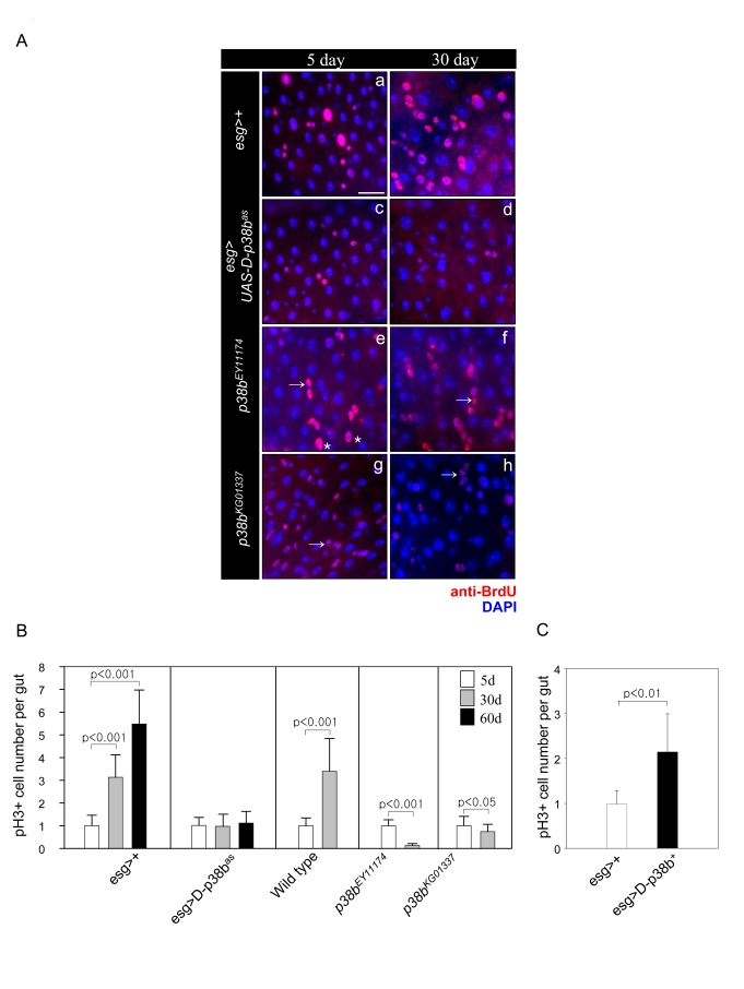
Figure 2.Effect of D-p38b MAPK signaling on DNA synthesis of intestinal cells and ISC division.
(A) Effects of D-p38b MAPK modulation on BrdU incorporation levels
in the adult midgut. Twenty-five day-old flies expressing esg>+ (a
and b), esg>UAS-D-p38bas(c and b), p38bEY11174
(e and f) or p38bKG01337 (g and h) were fed on
0.2 mg/ml BrdU media for 4 days, and stained with anti-BrdU. Overlay (DAPI,
blue; anti-BrdU, red). Asterisk indicates enlarged EC nuclei. Arrow
indicates small ISC, EB or EE cell nuclei. Scale bar, 5 μM. Original
magnification is 400x. (B) Effect of D-p38b MAPK activity on the
number of PH3-positive cells within the adult gut. Number of PH3-positive
cells detected per midgut of 5-, 30- and 60-day-old esg>+ or
esg>UAS-D-p38bas flies and 3- and 30-day-old control
flies, p38bEY11174 or p38bKG01337. The
number of PH3-positive cells detected per midgut of 5-day-old flies was set
as 1. White bar, 5-day-old flies; gray bar, 30-day-old flies; black bar,
60-day-old flies. P-values were calculated using Student's t-test. (C)
Effect of D-p38b MAPK activation on the number of PH3-positive cells.
Number of PH3-positive cells in the midguts of 5-day-old flies carrying esg>+
or esg>UAS-D-p38b+ were analyzed. White bar, esg>+;
gray bar, esg>UAS-D-p38b+. The number of PH3-positive
cells detected per midgut of 5-day-old flies was set as 1. P-values were
calculated using Student's t-test.