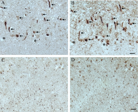
Figure 2. In another AD case, age 63, adjacent hippocampal
tissue sections demonstrate many of the AD-related pathological
structures (arrows) containing pMcm2 (A) are also positive
for hyper-phosphorylated tau (B) in the CA1 region.
Lower magnification of adjacent sections of the subiculum
shows the large number of NFT and plaques recognized by
pMcm2 (C) and AT8 (D). * denotes landmark vessel. Scale
bars= 50 μm (A,B), 100 μm (C,D).