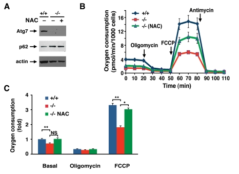
Figure 4.NAC treatment partially corrects the metabolic defect observed in Atg7 -/- MEFs.
(A) Western blot analysis of wild type (+/+) or Atg7-/-
MEFs for the expression of Atg7, p62 and actin (loading control) cultured
in the presence or absence on the antioxidant NAC (500 μM) for ten days. (B)
Primary wild-type and Atg7-/- MEFs that were cultured in the
absence or presence of 500 μM NAC for 10
days prior to cellular respiration measurement. Shown is a representative
tracing of oxygen consumption performed in triplicate under basal
conditions, following the addition of oligomycin (0.5 μM), the
pharmacological uncoupler FCCP (1 μM) or the Complex III inhibitor
antimycin A (0.25 μM). (C) Averaged metabolic profile from 4
separate experiments employing 3 independent primary isolates of WT and
Atg7-/- MEFs. Shown is the fold change +/- SEM in oxygen
consumption (WT MEF basal respiration =1) for WT MEFs and for Atg7-/-
MEFs that were cultured in the absence or presence of 500 μM NAC for 10
days prior to metabolic assessment.* p≤0.05; ** p≤0.01; NS= not
significant.