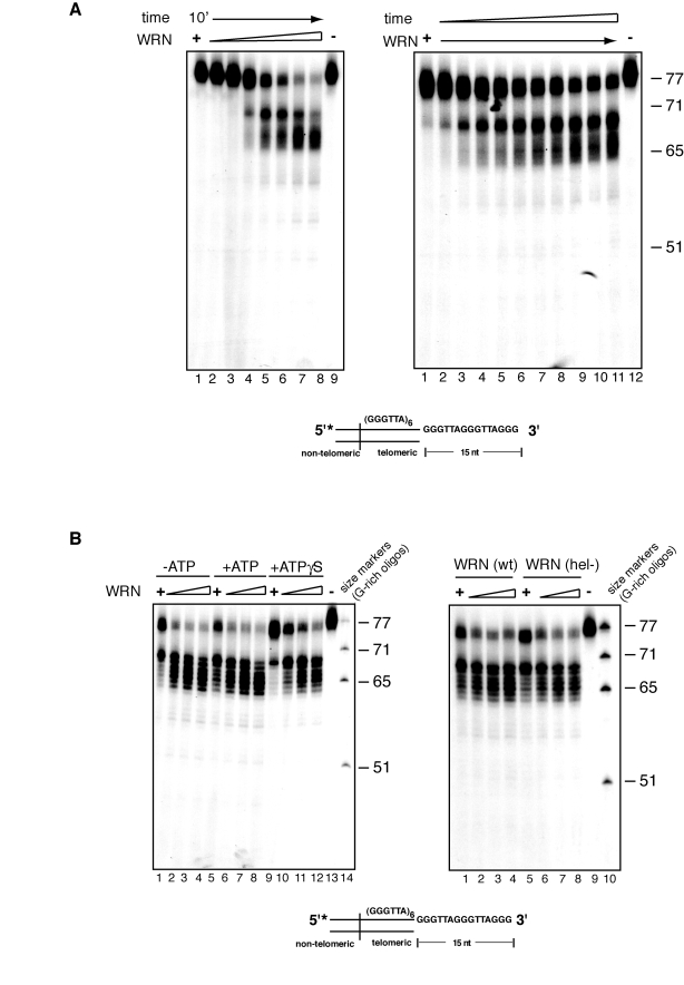
Figure 2.Concentration and time dependency of WRN exonuclease activity on telomeric substrates. (A) (left) 25 to 500 fmol of purified WRN were
incubated with 5'-32P-labeled, 3'-overhang telomeric DNA
substrate at 37°C for 10 min. The reaction products were analyzed by 12%
polyacrylamide-urea denaturing gel and autoradiography (lane 1 to 10, 25,
50, 100, 150, 200, 250, 300, 350, 400, 500 fmol of purified WRN; lane 11, DNA
substrate. (Right) 200 fmol of purified WRN was incubated with 5'-32P-labeled,
3'-overhang telomeric DNA substrate at 37°C from 0 to 10 min. The reaction
products were analyzed by 12% polyacrylamide-urea denaturing gel and
autoradiography (lane 1 to 11, 0, 1, 2, 3, 4, 5, 6, 7, 8, 9 and 10 min;
lane 12, DNA substrate. (B) (left) 100 to 400 fmol of
purified WRN were incubated with 5' 32P-labeled, 3'-overhang
telomeric DNA substrate in the absence or presence of 1.0 mM ATP or 1.0 mM
Adenosine 5'-[γ-thio]triphosphate
(ATPγS) at 37°C for 10 min. The
reaction products were resolved by 12% polyacrylamide-urea denaturing gel
and visualized by autoradiography (lane 1 to 4, 100, 200, 300, and 400 fmol
of WRN without ATP; lane 5 to 8, 100, 200, 300, and 400 fmol of WRN in the
presence of ATP, lane 9 to 12, 100, 200, 300, and 400 fmol of WRN in the
presence of 1.0 mM ATPγS; lane 13, DNA substrate; lane
14, (TTAGGG) repeats molecular size markers. (Right) 100 to 400 fmol
of purified WRN or WRN helicase mutant (K577M) were incubated with
telomeric DNA substrates at 37°C for 10 min. The reaction products were
analyzed by 12% polyacrylamide-urea denaturing gel and autoradiography
(lane 1 to 4, 100, 200, 300, and 400 fmol of WRN; lane 5 to 8, 100, 200,
300, and 400 fmol of helicase mutant WRN; lane 9, DNA substrate; lane 10,
(TTAGGG) repeats molecular size markers.