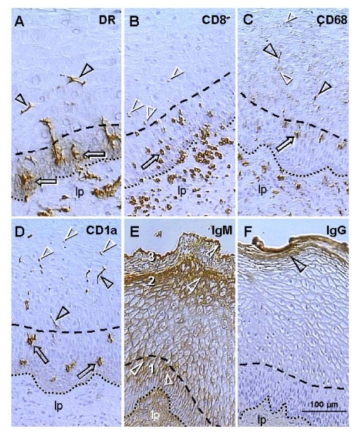
Figure 3.Uterine ectocervix immunohistochemistry as indicated above columns. (A) Dendritic cell
(DC) precursors secrete HLA-DR among parabasal cells (arrows) and
differentiate into mature DC (arrowheads). (B) T cells migrate
through parabasal layer (arrow) to parabasal/intermediate interface (dashed
line) and show fragmentation after entering the intermediate layer
(arrowheads). (C) Transformation of DC precursors into mature DC at
the top of parabasal layer is associated with CD68 expression (arrow).
Mature DC (black arrowheads) secrete CD68 material in intermediate layer
accompanying mature (intermediate) and aged (superficial) epithelial cells
(white arrowheads). (D) CD1a is expressed by DC precursors (arrows)
and mature DC (black arrowheads). Mature DC (Langerhans' cells) undergo
fragmentation in the mid intermediate layer (white arrowheads). (E)
Strong IgM binding (arrowheads) in upper parabasal [1], upper intermediate
[2] and upper superficial layers [3]. (F) IgG binds to the entire
superficial layer. For abbreviations see Figure 2. Reprinted from Ref. [4], © Antonin Bukovsky.