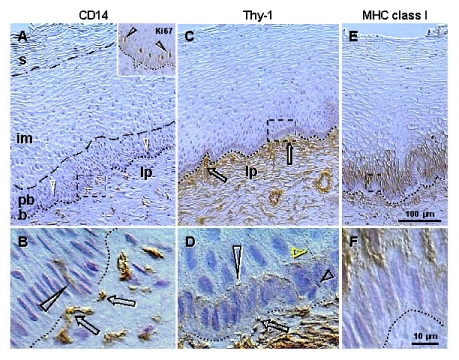
Figure 2.Peroxidase immunohistochemistry (brown color) of stratified epithelium of uterine ectocervix as indicated above columns and in the inset.
(A) CD14
primitive MDC in lamina propria (lp) associate with the epithelium basement
membrane (dotted line). Dashed box indicates detail shown in (B). b,
basal layer; pb, parabasal layer; im, intermediate layer; s,
superficial layer. Arrowheads, basal/parabasal interface; dashed line,
parabasal/intermediate interface; dashed/dotted line,
intermediate/superficial interface. Ki67 staining (inset) of epithelial
cells in lower parabasal layer (arrowheads). (B) CD14 MDC (arrows)
exhibit extensions among basal cells (arrowhead). (C) Pericytes of
microvasculature (arrows) associate with the basement membrane. (D)
Detail from (C) shows intercellular Thy-1 vesicles (arrow) secreted by
pericytes and migrating among basal cells (short black arrowhead) to
basal/parabasal interface (long
arrowhead). Yellow arrowhead indicates residual empty structures
("spikes"). (E) Strong MHC class I expression (W6/32
antibody specific for heavy chain) is characteristic of para-basal cells,
and diminishes in lower intermediate layers. Dashed box indicates detail
shown in (F). (F) Basal cells show no MHC class I expression.
Reprinted from Ref. [4], © Antonin Bukovsky.