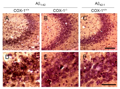
Figure 4.Increased oxidative damage in the hippocampus 7 d after Aβ 1-42 administration. Representative
photomicrographs of the CA1 and CA3 of the hippocampus from WT mice (A,
D) injected with Aβ1-42
that show numerous robustly nitrotyrosine-immunoreactive cells compared
with Aβ1-42-injected COX-1-/- mice (B,
E). Scale bar: A-C, 100 μm; D-F, 50 μm.