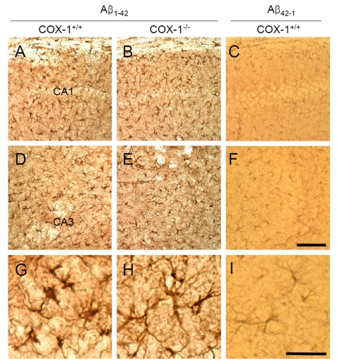
Figure 2.Increased astrocytic activation in the hippocampus 7 d after Aβ 1-42 administration. Representative
photomicrographs of the CA1 and CA3 of the hippocampus from WT mice (A,
D, G) injected with Aβ1-42 that shows numerous robustly
GFAP-immunoreactive astrocytes compared with Aβ1-42-injected
COX-1-/- mice (B, E, H). Scale
bar: A-F, 100 μm; G-I, 50 μm.