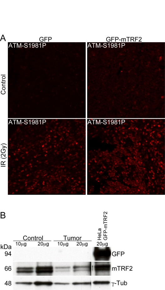
Figure 4.The ATM dependent damage response is not compromised in tumor samples.(A) Immunofluorescence
of ATM autophosphorylation after ionizing irradiation. Splenocytes were
irradiated with 5 Gy and microtome sections were prepared as described in
the "Materials and Methods" section. ATM activation was measured by
immunofluorescence with an antibody specific for the phosphorylation event
at serine 1981. The left panels represent cells from a control animal, the
right panel from an animal expressing the GFP-mTRF2 fusion. The upper
panels are before, the lower panels after irradiation. (B) Western
analysis of spleen from a secondary recipient mouse, which expressed
GFP-mTRF2 and died from a CD4/CD8+/+ T cell lymphoma. Protein samples were
probed with antibodies against GFP and mTRF2. g-Tubulin was included as a
loading control. As a positive control for GFP expression protein extract
from HeLa 1.2.11 cells expressing GFP-mTRF2 was loaded.