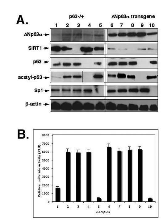
Figure 2.ShRNA silencing of ΔNp63-SIRT1-p53-Sp1 pathway. Mouse epidermal keratinocytes (2x10⁵ cells) from p63-/+
(samples 1-5) or overexpressing ΔNp63α(samples 6-10) were treated with control
media (samples 1 and 6), SIRT1 inhibitor (Sirtinol, 100 μg/ml for 24 h;
samples 2 and 7), or transfected with the SIRT1 shRNA (samples 3 and 8), p53
shRNA (samples 4 and 9), and sh-Sp1 RNA (samples 5 and 10).
(A) Immunoblotting with indicated antibodies (dilutions: anti-ΔNp63, 1:500;
anti-SIRT1, 1:300; anti-Sp1, 1:300; anti-p53, 1:500; anti-acetyl-p53,
1:400; anti-β-actin, 1:400). The vertical lines separate data obtained from
independent protein gels.
(B) mTERT promoter luciferase reporter assay.
Mouse keratinocytes (1.0 x 10⁵) were transfected with the pGL3-347-Luc
plasmid (0.5 μg) or the pGL3 control plasmid (0.5 μg) by using FuGENE6 transfection reagent
(Roche Diagnostics). 3 ng of the pRL-SV40 (Promega) was used as a
normalization control. Measurements were performed by using the Dual
Luciferase reporter assay system (Promega) and a BioOrbit 1251
luminometer. The activity of each TERT promoter fragment was expressed as
a relative value. All of the data (mean +SD) were from at least three
independent experiments.