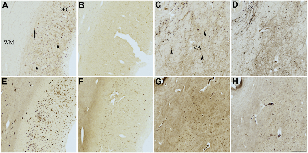
Figure 9.Effect of meclofenoxate hydrochloride (6) on histochemical staining of acetylcholinesterase (AChE) and butyrylcholinesterase (BChE). Representative photomicrographs of histochemical staining of AChE (A–D) and BChE (E–H). Staining at pH 6.8 demonstrates AChE- (A) and BChE (E)-associated plaques in the Alzheimer’s disease (AD) orbitofrontal cortex (arrows). Staining at pH 8.0 demonstrates AChE (C) and BChE (G) associated with normal neural structures in the AD thalamus (arrowheads showing neurons). Meclofenoxate hydrochloride (6) inhibits AChE (B) and, to a certain extent, BChE (F) associated with AD plaques but not AChE (D) and BChE (H) associated with normal neural elements. Note, for ease of reference, identical images of the positive control staining of AChE and BChE at pH 6.8 and 8.0 (A, C, E, G) were used herein and in Figures 4-8 (A, C, E, G) to help compare directly the effects of each senolytic or nootropic agent on the standard Karnovsky-Roots (KR) histochemical staining method. Abbreviations: OFC, orbitofrontal cortex; VA, ventroanterior thalamic nucleus; WM, white matter. Scale bar = 500 μm.