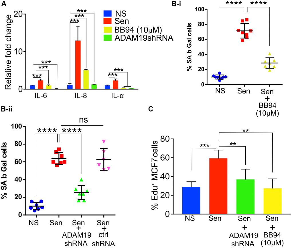
Figure 3.ADAM19 knockdown and pharmacological inhibition reduced senescence in human fibroblast cells caused by irradiation. (A) The mean fold change in the expression levels of senescence-associated secretory phenotype (SASP) cytokines IL-6, IL-8, and IL-1α was measured by qRT-PCRIMR-90 cells ten days after exposure to 100 Gy X-ray. The expression levels were quantified relative to untreated control samples and normalized to the housekeeping gene actin (ACTB) to control for differences in input RNA and reverse transcription efficiency. Data are presented as mean ± SEM from three independent experiments. The results represent the relative fold change in gene expression compared to control. Increased expression of SASP cytokines, such as IL-6, IL-8, and IL-1α, indicates a senescence response and may reflect the inflammatory environment of irradiation-induced cellular damage, which was significantly reduced in cells with ADAM19 knockdown or inhibition following treatment with BB-94. (B-i) IMR-90 cells were subjected to SA β-Gal staining ten days after irradiation (10Gy), following 10 µM BB-94 treatment. (B-ii) IMR-90 cells were subjected to SA β-Gal staining ten days after irradiation (10Gy), following transduction with either ADAM19 or scrambled shRNA. The percentage of SA β-Gal positive cells was quantified and presented as mean ± SEM, with three independent experiments with two technical replicates per group. (C) MCF-7 cells were treated with conditioned media collected from IMR-90 cells ten days after irradiation with or without ADAM19 shRNA or with or without Batimastat (10 µM). Results plotted as mean % EdU positive cells. (A–C) Error bars indicate SEM. The significance level was determined by ANOVA and denoted by asterisks: *p < 0.05, **p < 0.01, ***p < 0.001, and ****p < 0.0001. In (A, B-i, B-ii and C) “NS” represent non senescence and ‘Sen’ represent senescence.