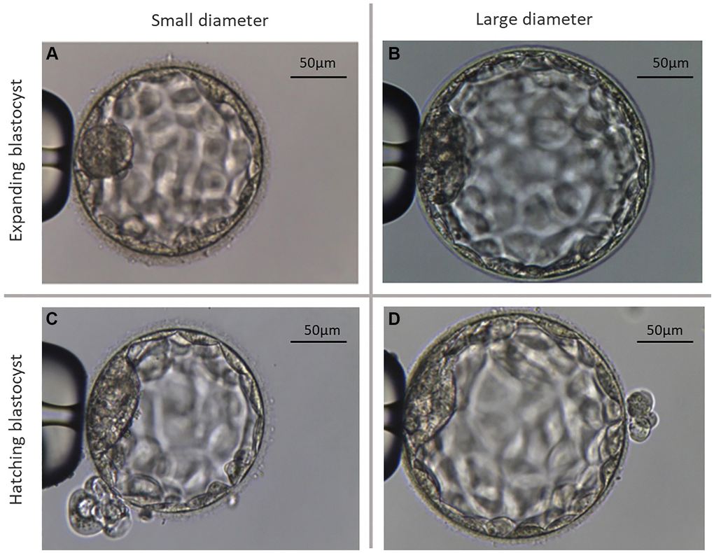
Figure 1.Representative microscopic photographs of the two different types of analyzed blastocysts. (A) Shows an exemplary photograph of a large expanding blastocyst (group Bl. 4; ≤185 µm). (B) Shows an exemplary photograph of a large expanding blastocyst (group Bl. 4; >185 µm). (C) Shows a small hatching blastocyst (group Bl. 5; ≤195 µm). (D) Shows a large hatching blastocyst (group Bl. 5; >195 µm).