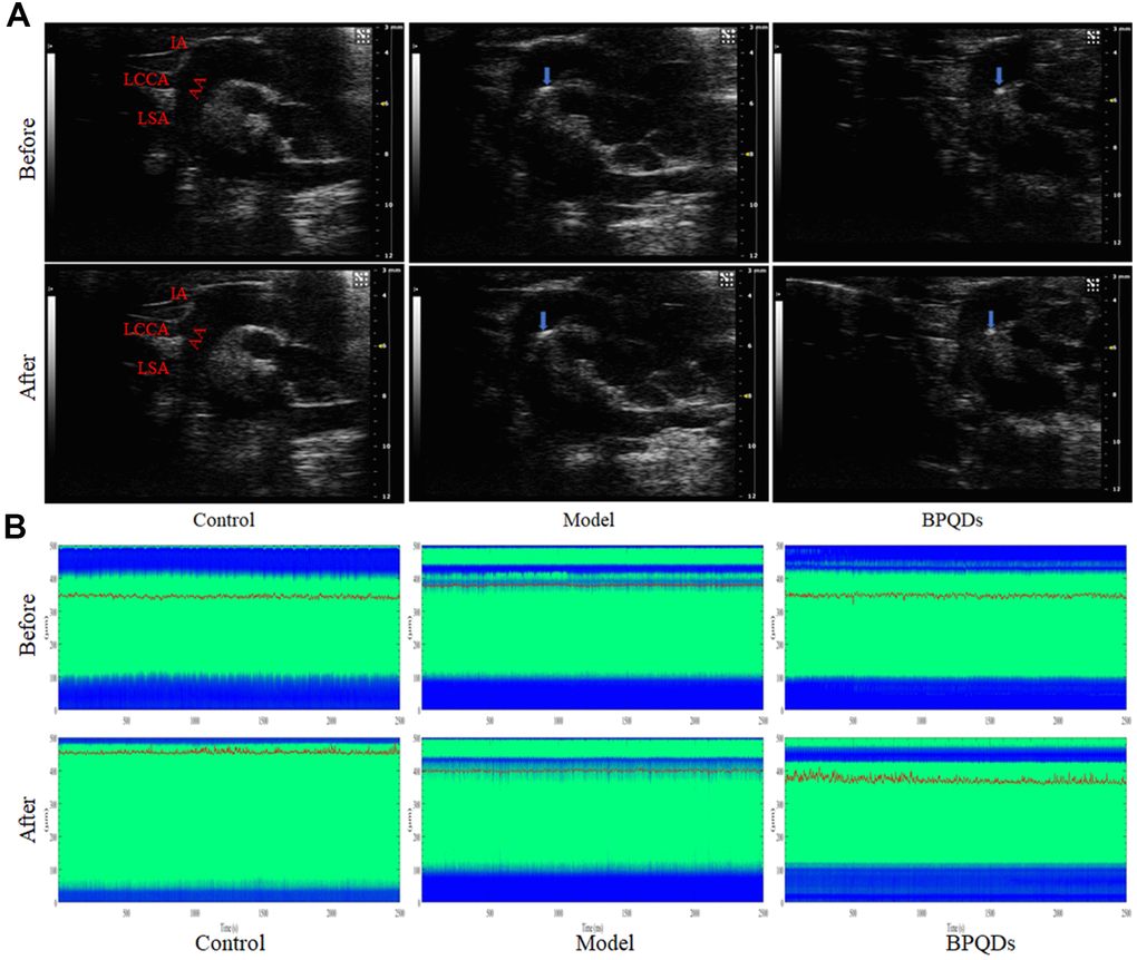
Figure 7.(A) Ultrasonography of the aortic plaque (blue arrow) before and after treatment. AA: Aortic arch, IA: Innominate artery, LCCA: Left common carotid artery, LSA: Left subclavian artery. (B) Comparison of photoacoustic microscopy images of abdominal aorta in mice after treatment. (Before: before the injection of vasodilator, After: after the injection of vasodilator, the green area between the blue bands indicates the internal diameter of the abdominal aorta, the red curve shows the change of the internal diameter of the abdominal aorta over time, and the corresponding ordinate is the internal diameter of the abdominal aorta).