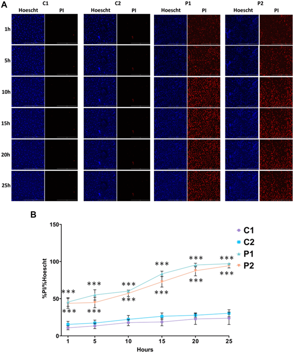
Figure 8.Sensitivity to ferroptosis in control (C1, C2) and MS (P1, P2) cells. Cells were treated with 5 μM erastin and stained with Hoechst (blue fluorescence) and propidium iodide (PI, red fluorescence) to distinguish between dead and live cells. (A) Representative images of live and dead cells upon addition of erastin for 25 hours. Scale bar = 20 μm. (B) Quantification of cell death over time. Data represent the mean ± SD of three independent experiments. ***p-value < 0.0001 between control and MS fibroblasts.