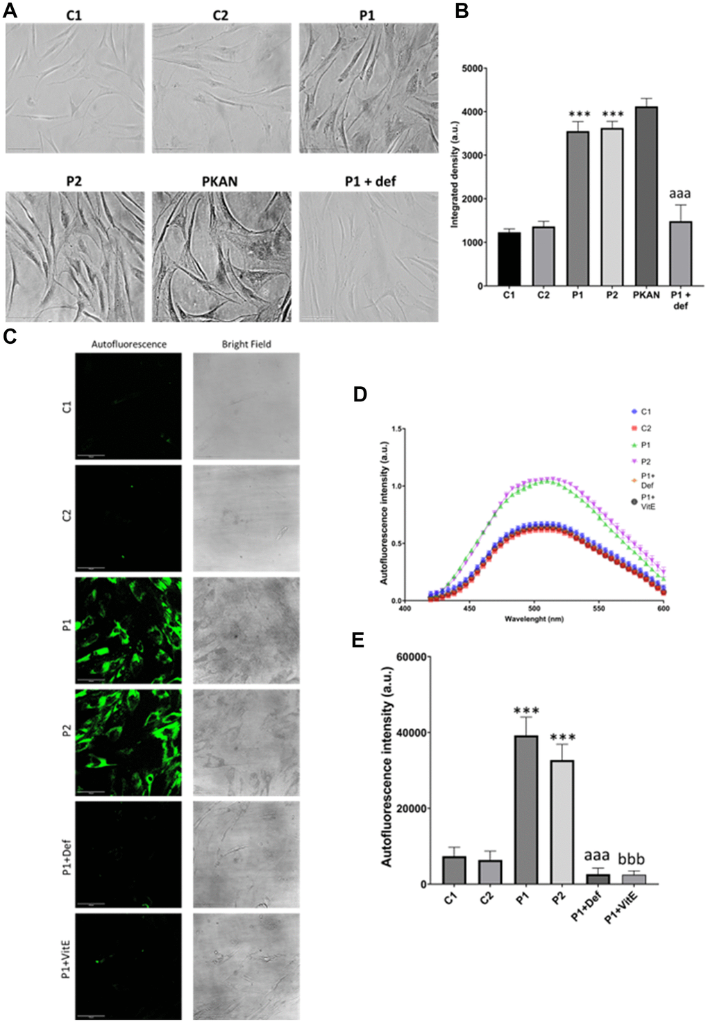
Figure 5.Lipofuscin accumulation in control (C1, C2) and MS (P1, P2) fibroblasts. P1 cells were treated with 100 μM deferiprone (P1 + def) to confirm the dependence of lipofuscin on iron. P1 cells were treated with 50 μM vitamin E (P1 + VitE) to confirm the dependence of lipofuscin accumulation on lipid peroxidation. (A) Representative images of Sudan Black staining. Scale bar = 20 μm. (B) Quantification of integrated density. (C) Representative autofluorescence and bright field images. Scale bar = 20 μm. (D) Autofluorescence spectra of lipofuscin granules measured by confocal laser scanning microscopy. (E) Quantification of autofluorescence intensity. Data represent the mean ± SD of three independent experiments. ***p-value < 0.0001 between control and MS cells. aaap-value < 0.0001 between untreated and deferiprone-treated P1 cells. bbbp-value < 0.0001 between untreated and vitamin E-treated P1 cells. a.u.: arbitrary units.