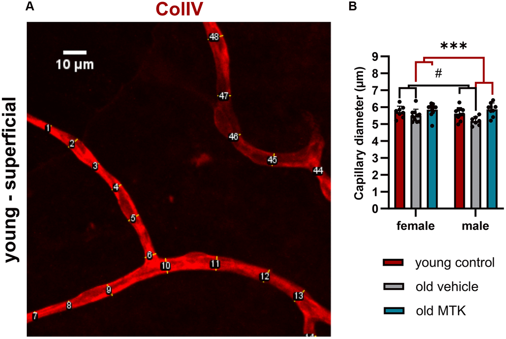
Figure 4.Capillary diameter in superficial retinal layers of young untreated, vehicle-treated and MTK-treated old mice. The capillary diameter was measured using ImageJ. (A) Representative image showing how the capillary diameter was measured. (B) Capillary diameter in the superficial retinal layers of young untreated, vehicle-treated and MTK-treated old mice. The values are represented as bar graphs and scatter plots ± SDs, n = 7–11. Two-way ANOVA (main factors: group and sex) followed by a Dunnett multiple comparison test. #p < 0.05 for the young control vs. the old vehicle-treated group; ***p < 0.001 for the old MTK-treated group vs. the old vehicle-treated group.