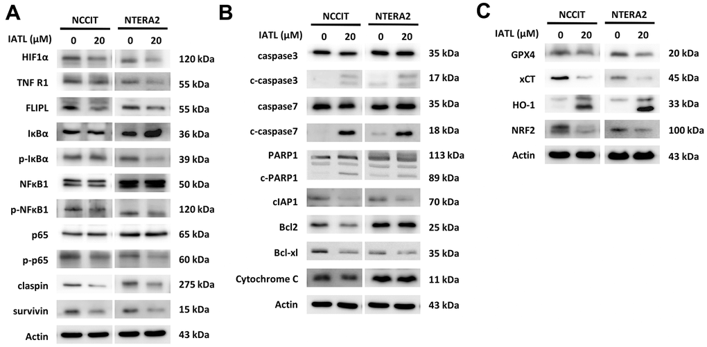
Figure 3.IATL triggered apoptosis and ferroptosis pathways in testicular cancer cells. (A) The western blot implied the decreasing expression of p-IκBα, p-NFκBα1 and p65, the downstream molecules of the TNFR1 pathway. (B) The apoptosis-related proteins presented the expected trends after IATL treatment. Our hypothesis that IATL could induce apoptosis in testicular cancer cell lines has been confirmed. (C) GPX4, xCT, and NRF2 demonstrated declining trends, and HO-1 presented an increasing trend. The results verified that IATL is related to ferroptosis.