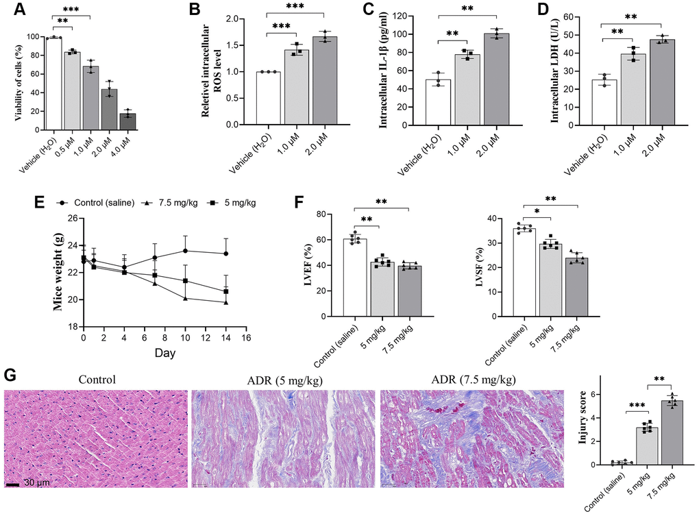
Figure 1.Adriamycin (ADR) treatment damages cardiomyocyte and impaired cardiac function of C57BL/6 mice. (A) Cardiomyocytes were treated by a serial of ADR, and ADR decreases the viability of cardiomyocytes; (B–D) ADR stimulates oxidative stress and inflammatory response. ADR causes increasing of intracellular reactive oxygen species (ROS) (A) and interleukin-1β (IL-β) (B) lactate dehydrogenase (LDH) (C) level, and LDH levels in primary cardiomyocytes (D); (E) ADR causes obvious decreasing of body weight, and impaired left ventricular ejection fraction (LVEF) and left ventricular systolic function (LVSF) in mice (F); (G) Hematoxylin and Eosin (H&E) staining in ADR-treated myocardium of mice. Increased collagen between muscle, irregular arrangement of myocardial cell nuclei, granular and interstitial hemorrhage could be observed. **p < 0.01, ***p < 0.001.