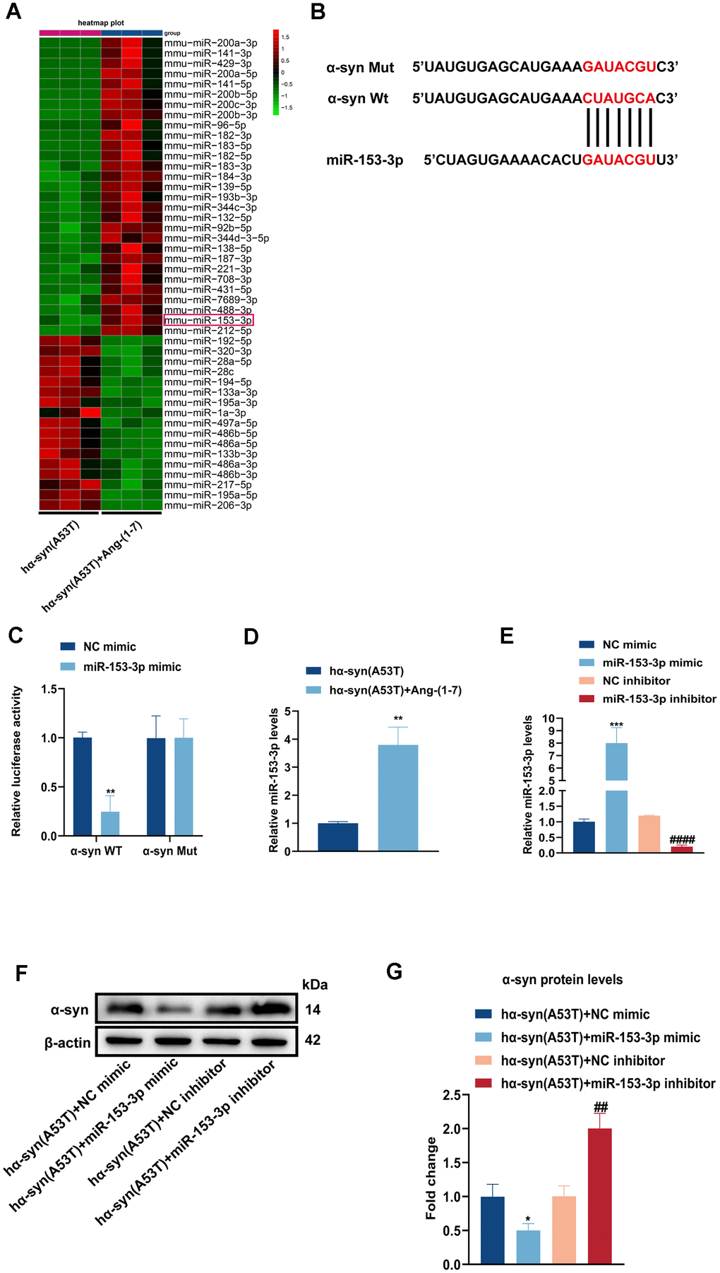
Figure 2.Overexpression of miR-153-3p reduces the level of α-syn and relieves the apoptosis in the hα-syn(A53T) overexpressed dopaminergic neurons. (A) The heatmap showed differentially expressed miRNAs in right SN of mice for every group (n = 3). (B) Probable binding site of miR-153-3p to α-syn. (C) The luciferase reporter assay to verify the combination between miR-153-3p and α-syn (n = 3). (D) The miR-153-3p level in SN of each mice group were assessed by qRT-PCR (n = 3). (E) The miR-153-3p levels of primary dopaminergic neurons in indicated groups were analysed by qRT-PCR (n = 3). (F) The α-syn level of primary dopaminergic neurons in each group was tested by Western blot (n = 3). (G) Quantitative evaluation of α-syn level.
(H) Cell cytotoxicity of primary dopaminergic neurons in each group was tested by LDH assay (n = 3). (I) The levels of Bcl2, Bax and cleaved caspase-3 in every group were observed by Western blot (n = 3). (J) Quantitative evaluation of Bcl2 level. (K) Quantitative evaluation of Bax level. (L) Quantitative evaluation of Cleaved caspase-3 level. Data are shown as the mean ± SD. *P<0.05 versus the hα-syn(A53T) + NC mimic group; **P<0.01 versus the NC mimic, hα-syn(A53T) or hα-syn(A53T) + NC mimic group; ***P<0.001 versus the NC mimic or hα-syn(A53T) + NC mimic group; #P<0.05, ##P<0.01 and ###P<0.001 versus the hα-syn(A53T) + NC inhibitor group. ####P<0.0001 versus the NC inhibitor group.