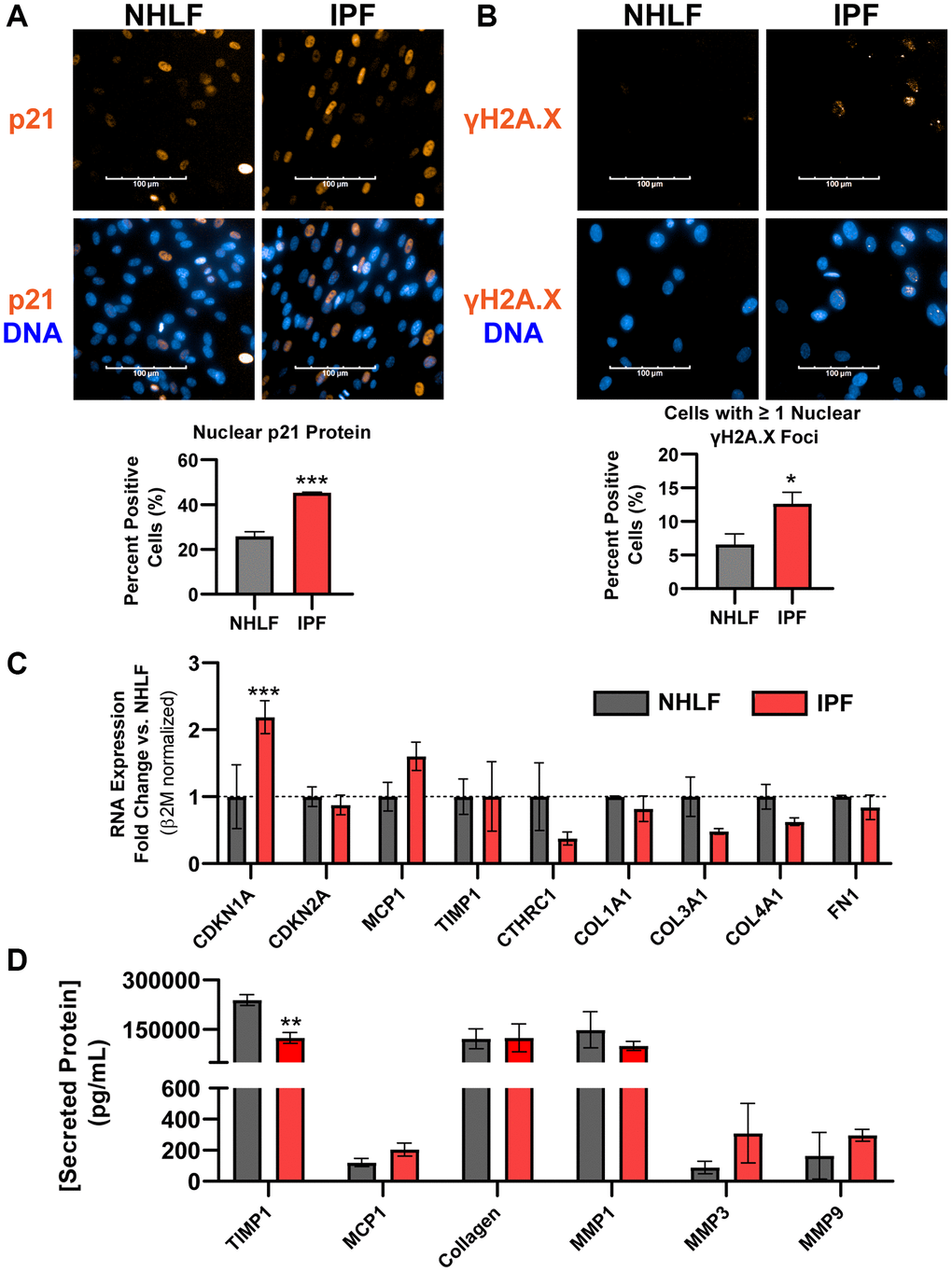
Figure 2.DNA damage is a hallmark of commercial primary IPF fibroblasts in vitro, but not Cdkn2a/p16ink4a expression or a fibrotic secretome. (A) Primary NHLF and IPF cells (n = 3 different donors, each with 2 technical replicates) were cultured at the same density overnight on standard tissue culture plates in low-serum growth medium and were subsequently fixed. Immunocytochemistry was performed to fluorescently label nuclei (blue) and p21Waf1/Cip1 (orange). Images were acquired with a 40X water objective using an Operetta High Content Screening instrument and intensity of nuclear p21Waf1/Cip1 was quantified and calculated as a positive percentage of each population. (B) NHLF and IPF cells were treated and imaged as in (A), and immunocytochemistry was performed to fluorescently label nuclei (blue) and DNA damage via nuclear γH2A.X (Ser139) foci (orange). The number of nuclear foci per cell was quantified and cells with one or more were reported as positive. (C) Following overnight culture in standard tissue culture plates, NHLF and IPF cells were lysed, and qPCR was performed. Expression of senescence and fibrosis-related matrix and secreted factor genes was assessed, and data were normalized to β2m housekeeper expression using the 2−ΔΔCt method versus NHLF cells. (D) Following overnight culture as in (C), cell culture supernatants were collected and assayed for determination of the concentration of common fibrosis-related secreted proteins by MSD kits. Statistical analysis was performed using an unpaired t-test (A, B) or a two-way ANOVA with a Bonferroni post-test (C, D) in GraphPad Prism: *p < 0.05, **p < 0.01, ***p < 0.001. Error bars represent standard error of the mean (SEM).