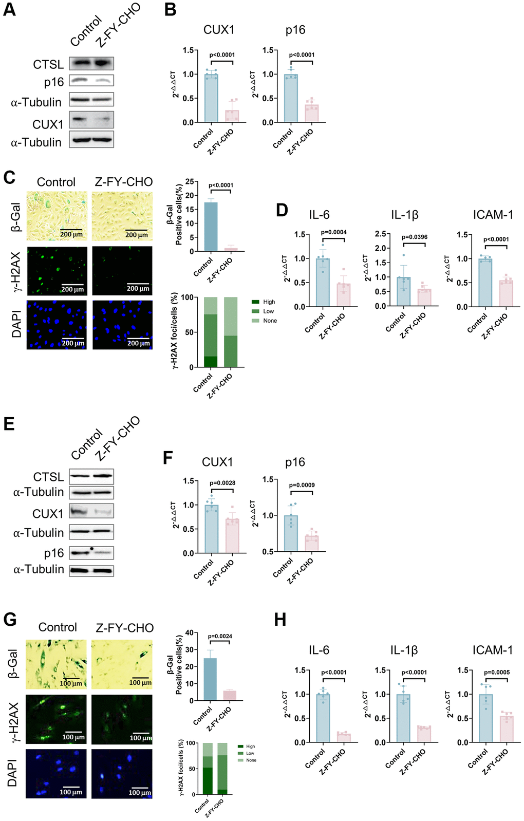
Figure 3.Inhibition of CTSL protease activity decreases the expression of CUX1 and p16INK4a, and inhibits cellular senescence in human ECs and VSMCs. (A, B) Western blot and qPCR analyses showing that inhibition of CTSL protease activity by Z-FY-CHO downregulates CUX1 and p16INK4a in human ECs. (C, D) inhibition of CTSL protease activity by Z-FY-CHO suppresses cellular senescence in human ECs as evidenced by SA-β-gal and γ-H2AX staining as well as by the expression of the SASP genes IL6, IL-1β, and ICAM-1. Quantitative plots for both β-gal+ cells (%) in SA-β-gal staining and γ-H2AX foci/cells (%) with γ-H2AX staining were shown on the right side of the panel C. DAPI staining visualizes the presence of nuclei as a control for the cells in this analysis. (E, F) Western blot and qPCR analyses showing that inhibition of CTSL protease activity by Z-FY-CHO downregulates CUX1 and p16INK4a in human VSMCs. (G, H) inhibition of CTSL protease activity by Z-FY-CHO suppresses cellular senescence in human VSMCs as evidenced by SA-β-gal and γ-H2AX staining as well as by the expression of the SASP genes IL6, IL-1β, and ICAM-1. Quantitative plots for both β-gal+ cells (%) in SA-β-gal staining and γ-H2AX foci/cells (%) with γ-H2AX staining were shown on the right side of the panel G. DAPI staining visualizes the presence of nuclei as a control for the cells in this analysis. Data for Western blots represent three biologically independent samples (n = 3). Data for qPCR represent a combination of three (n = 3) biologically independent experiments. Data for both SA-β-gal staining and γ-H2AX staining represent three biologically independent samples (n = 3).