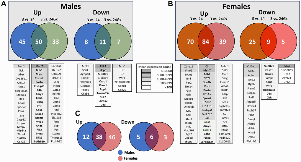
Figure 5.Transcriptomic changes in the left ventricle induced by aging and gonadectomy. Differentially expressed genes (DEGs, |Log2 fold change| ≥1) between 3-month-old (3 m) and 24-month-old males (24 m) (A) or between 3-month-old and 24-month-old female (B) mice, intact or Gx. (A) Venn diagrams showing DEGs specific to aging intact or Gx males as well as shared genes either up (left) or down-regulated (right). Venn diagrams showing DEGs specific to aging intact or Gx females and common genes either up (left) or down-regulated (right). Below the Venn diagrams are lists of up to twenty genes ranked by their mean expression levels for each comparison from highest to least expressed. Legend for the background colouring of the gene list based on mean expression count is illustrated. In boldface are listed genes that are common between males and females. (C) Venn diagrams for the common upregulated genes (left) and downregulated genes (right) by aging between males and females. Only genes having a mean normalized count over 50 from RNA-Seq data were considered.