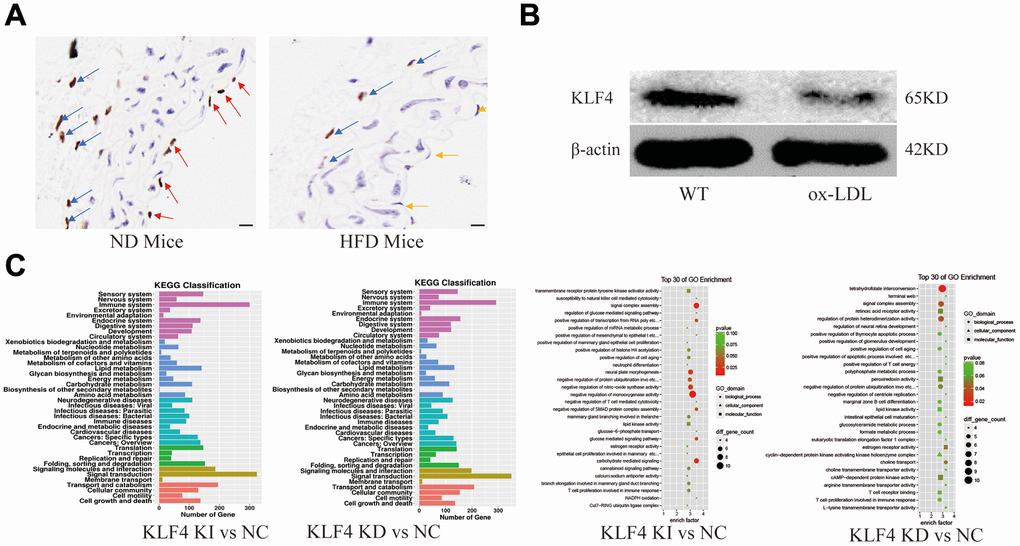
Figure 1.Analysis of KLF4 protein expression and EC dysfunction. (A) Immunohistochemical detection of vascular wall KLF4 expression. Scale bar, 20 μm. Representative images (n=5) are shown. Red arrow, KLF4 expression in the intima of mice fed a normal diet. Yellow arrow, intima lacking KLF4 expression in mice fed a high-fat diet. Blue arrow, KLF4 expression in adventitial fibroblasts. (B) Western blotting analysis of KLF4 protein expression in cultured HUVECs (100 μg/ml ox-LDL, 24 h) (n=5). (C) GO and KEGG analysis of the pathways after KLF4 knock in and knock out in HUVECs.