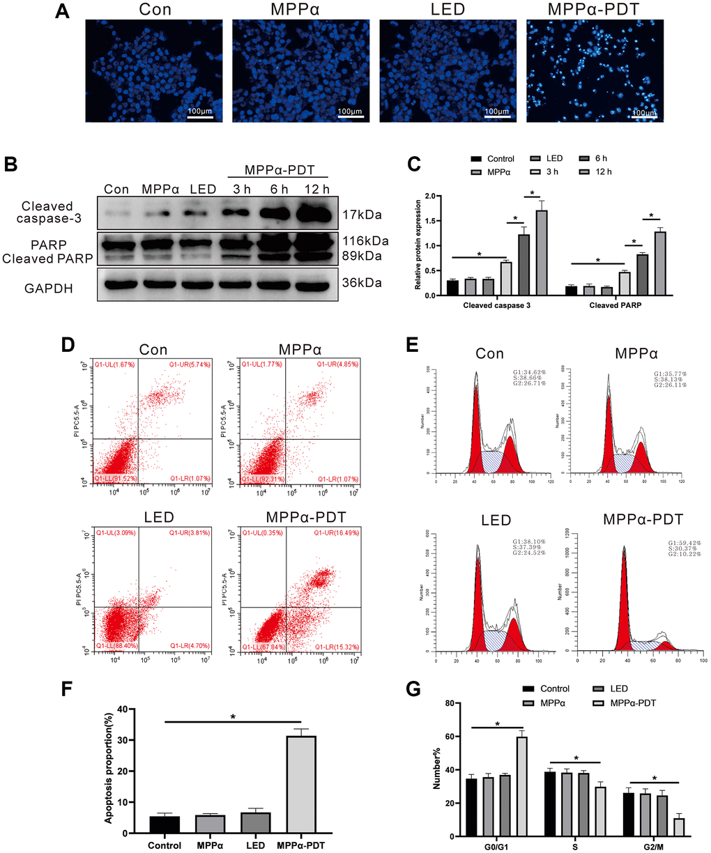
Figure 1.MPPα-PDT induces HOS cells cycle arrest and apoptosis. (A) HOS cells nuclei were examined for apoptotic morphological changes under a fluorescence microscope (magnification: ×200) after MPPα-PDT treatment for 12 h. (B, C) Cells were harvested after MPPα-PDT treatment for 3, 6, and 12 h and western blot used to evaluate cleaved caspase-3 and cleaved PARP levels. (D–F) Apoptosis was determined by flow cytometry. (E–G) Cell cycle distribution was analyzed using flow cytometry. *P < 0.05 vs. control group. All data represent the mean ± SD of 3 independent experiments.