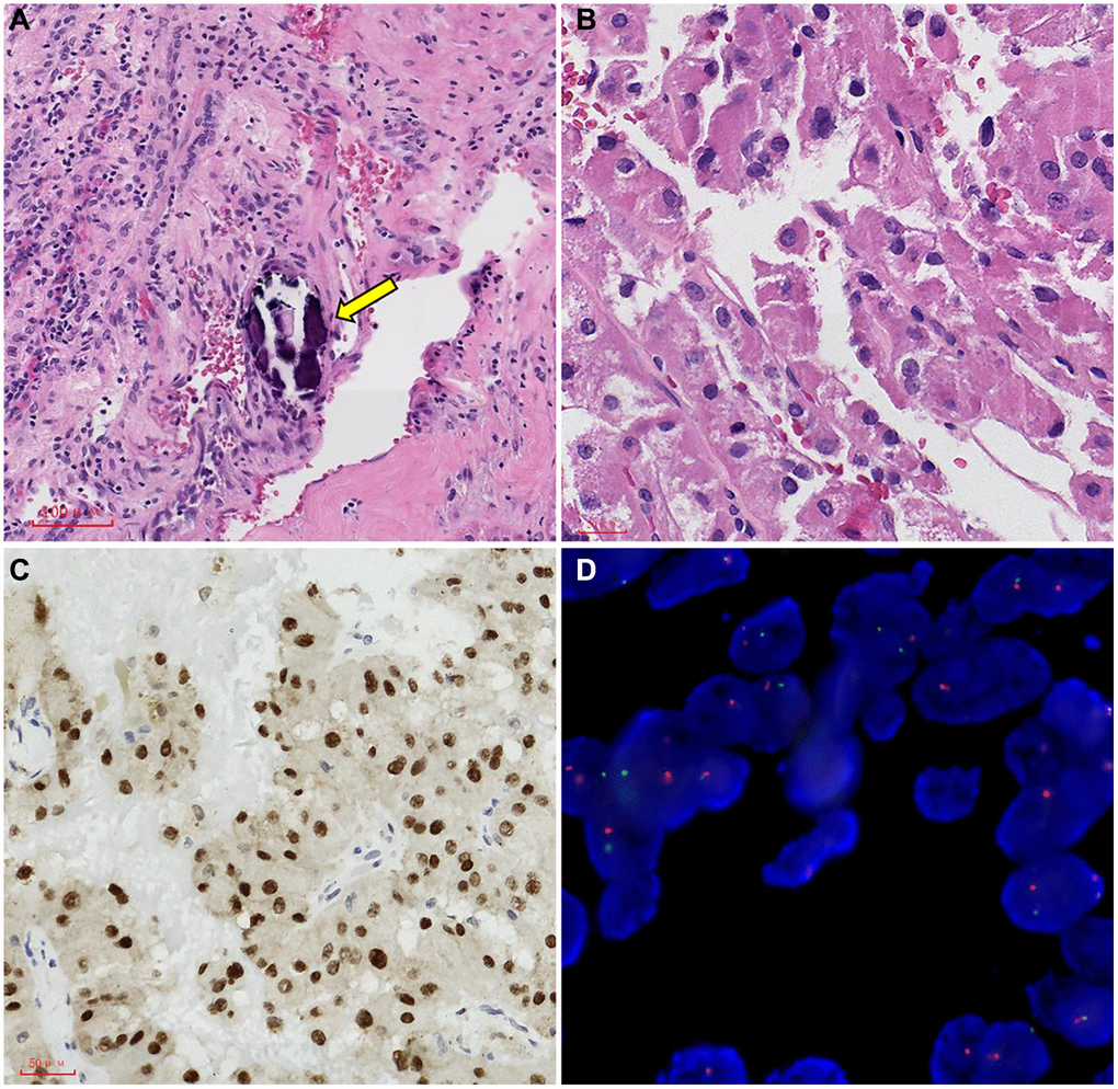
Figure 5.Representative images of TFE3 immunohistochemical staining and microscopic appearance for Xp11.2 RCC. (A) Blood sinusoid and Psammoma bodies were abundant in intercellular substance. The arrow points to Psammoma bodies; (B) abundant and deeply stained eosinophile cytoplasm, similar to renal clear cell carcinoma; (C) the results of immunohistochemistry showed that TFE3 was strongly positive in cancerous tissue; (D) FISH test results: 100 cells were counted, and the number of cells with TFE3 gene breakage was more than 20. TFE3 gene probe: Broken (positive).