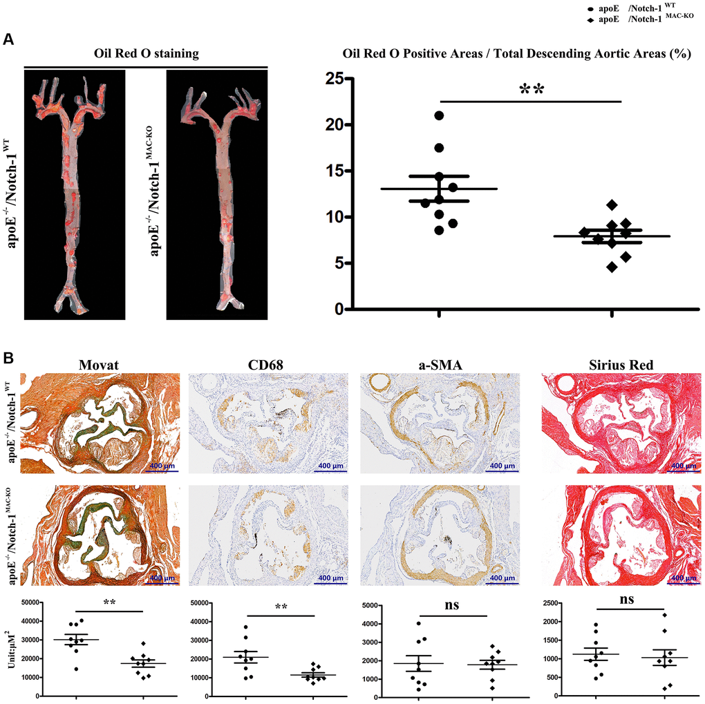
Figure 2.Specific deletion of Notch-1 in macrophage repressed AS. (A) Oil red O staining in thoracic aortas and quantitative analysis revealed that Notch-1MAC-KO significantly decreased the atherosclerotic plaques compared with Notch-1WT. **P < 0.01: ApoE−/−/Notch-1MAC-KO vs. ApoE−/−/Notch-1WT. (B) Movat, α-SMA, CD68 and Sirius red staining and quantitative analysis revealed that Notch-1MAC-KO could decrease plaque cellularity (Movat staining) and macrophage infiltration (CD 68) compared with Notch-1WT. However, there was no significant difference in smooth muscle cell content (α-SMA staining) and fibrotic lesions (Sirius red staining) between the two groups. **P < 0.01: ApoE−/−/Notch-1MAC-KO vs. ApoE−/−/Notch-1WT.