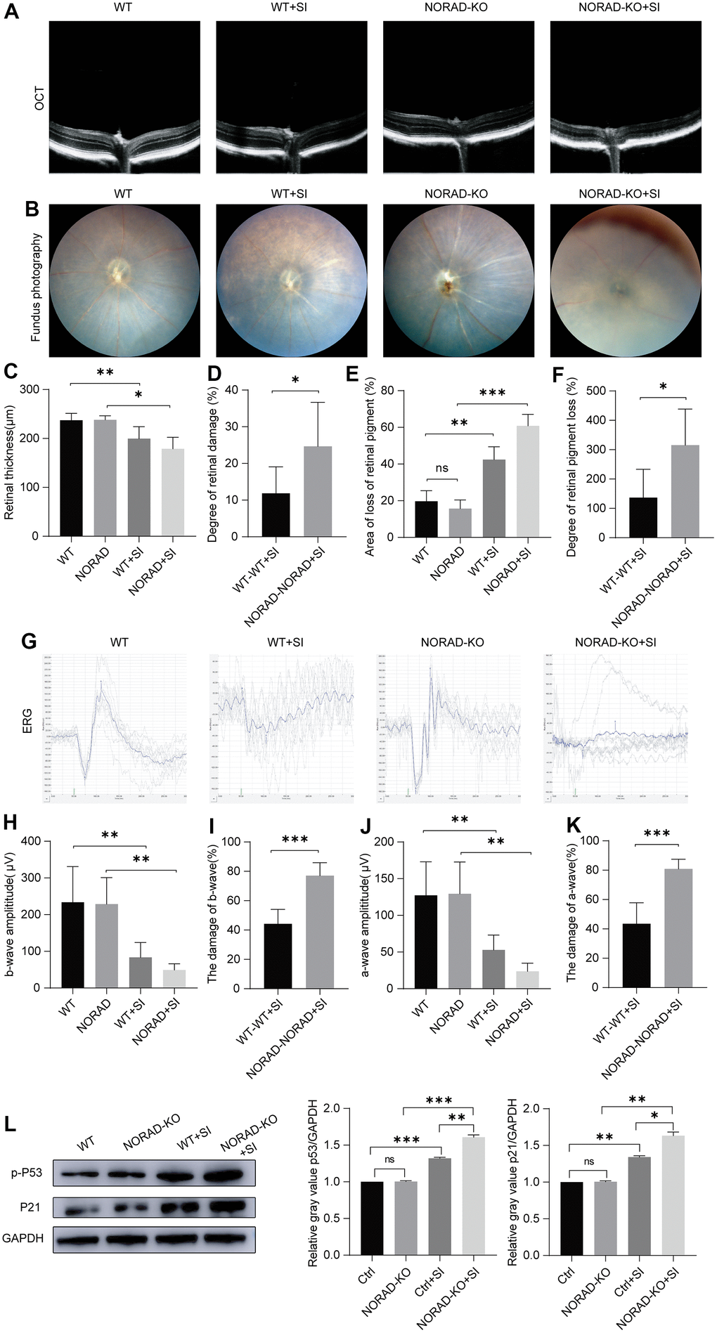
Figure 3.Injury effects of NORAD knockout on the retina of NaIO3-treated mice. Different techniques were used to detect retinal damage in wild-type mice and NORAD knockout mice treated with sodium iodate. (A) Optical coherence tomography (OCT) was performed seven days after NaIO3 treatment for all three study groups. (B) Funduscopic examinations were performed seven days after SI injection. Representative OCT and fundus fluorescence shows the RPE of the mice in each experimental group. (C) Statistical graph of retinal thickness. (D) Statistical graph of retinal damage between WT mouse and NORAD-KO mouse after SI injection. (E) Statistical graph of retinal pigment loss area. (F) Statistical graph of retinal pigment loss degree between WT mouse and NORAD-knockout mouse after SI injection. (G) Electroretinography was performed to investigate the function of the retina in response to light. (H) Statistical graph of b wave amplitude change. (I) Statistical graph of the degree of change in b wave amplitude between WT mouse and NORAD-knockout mouse in eight days after SI injection. (J) Statistical graph of a wave amplitude change. (K) Statistical graph of the degree of change in a wave amplitude between WT mouse and NORAD-knockout mouse in eight days after SI injection. n≥3, *P < 0.05, **P < 0.01, ***P < 0.001. (L) Western blot was used to detect the p-P53 and P21 expression levels. n = 3, *P < 0.05, **P < 0.01, ***P < 0.001.